


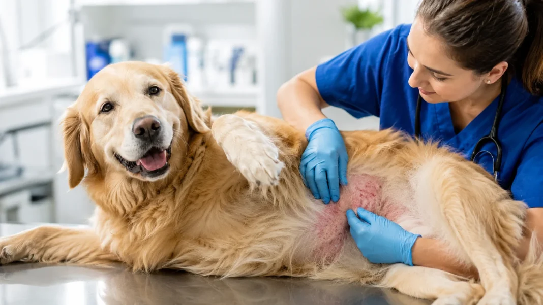
Dog Skin Conditions: Pictures, Causes and Treatments
Dog skin conditions can appear as red patches, bald spots, bumps, scabs, flaky skin, sores, dark pigmentation, greasy fur, or areas that your dog constantly licks and scratches. Unfortunately, several very different diseases can look almost identical in photographs, which means identifying the appearance is only the first step toward finding the cause. Allergies, fleas, mites, bacterial infections, yeast, ringworm, hot spots, and underlying medical disorders are among the possibilities. Some problems remain mild and localized, while others spread quickly or require prescription treatment. This guide explains common dog skin conditions, what they can look like in pictures, likely causes, typical treatments, diagnostic tests, safe home care, prevention, and signs that mean your dog should see a veterinarian.
What Are Dog Skin Conditions?
A dog skin condition is any disorder affecting the skin, coat, hair follicles, claws, or structures associated with the skin. Veterinarians commonly use the broad term dermatitis when inflammation is present but the exact cause has not yet been identified. Skin disease can produce itching, redness, scaling, hair loss, odor, oiliness, crusting, pigmentation changes, bumps, or discharge. A dog may have one small lesion or extensive changes across much of the body. Because the skin has a limited number of ways to respond to irritation, very different diseases can create remarkably similar appearances. That overlap is why photographs are helpful for recognizing abnormalities but rarely provide a complete diagnosis.
Itching, medically called pruritus, is one of the most important clues when evaluating canine skin disease. A dog may scratch with its hind legs, lick its paws, chew at the tail, rub its face on furniture, or repeatedly roll and bite irritated areas. Excessive self-trauma can eventually create more visible damage than the original condition. For example, an allergic dog may initially have subtle redness, but repeated scratching can cause broken hairs, scabs, bleeding, and secondary infection. Some diseases, however, cause substantial hair loss with relatively little itching. Veterinarians therefore ask not only what the skin looks like but also how intensely the dog scratches and whether itching appeared before or after the visible lesions.
Acute and chronic skin disease can look quite different. A newly irritated area may appear bright red, warm, moist, or covered with small bumps, while skin affected for months can become thick, dark, rough, and heavily pigmented. Merck notes that chronic dermatitis can eventually produce thickening, color changes, scaling, discharge, and increased dryness, sometimes making the original problem difficult to recognize. Secondary bacterial or yeast infections commonly complicate long-standing inflammation and may become major sources of itching themselves. This means a dog can have several problems at once, such as environmental allergy combined with yeast overgrowth and bacterial pyoderma. Successful treatment often requires addressing every layer rather than choosing one diagnosis from appearance alone.
A dog’s age, breed, season, environment, parasite prevention, and health history can significantly change which skin conditions are most likely. Atopic dermatitis commonly begins relatively early in life, while endocrine disorders are more frequently investigated in older dogs with recurring hair or skin changes. Puppies can develop conditions such as demodicosis, superficial bacterial infections, and ringworm that may create similar patches of hair loss. Dogs with heavy skin folds are more vulnerable to problems in warm, moist areas, while outdoor exposure can increase contact with fleas, mites, insects, vegetation, or fungal spores. No single factor confirms a diagnosis, but combining these clues helps veterinarians narrow a long list of possible causes.
Skin disease deserves attention because the skin is not simply cosmetic. It protects the body from microorganisms, prevents excessive water loss, participates in temperature regulation, and forms a major barrier between the dog and its environment. When that barrier becomes damaged, microorganisms that normally live harmlessly on the skin may multiply and cause secondary problems. Repeated scratching can also interfere with sleep, activity, appetite, and overall quality of life. Severe infections may become painful rather than merely itchy, and some skin conditions can signal diseases affecting other parts of the body. An ongoing, expanding, painful, or frequently recurring problem therefore deserves veterinary evaluation even if the dog otherwise seems healthy.
Dog Skin Conditions Pictures: What Common Problems Look Like
Flea allergy dermatitis often appears around the lower back, rump, base of the tail, and backs of the thighs. Pictures may show thinning fur, patchy baldness, red skin, small crusted bumps, scratches, or scabs concentrated toward the rear of the dog. In long-standing cases, skin can become darker and thicker from repeated inflammation and chewing. Importantly, owners may never see a large number of fleas because a sensitive dog can react strongly to relatively limited exposure and may remove fleas through constant grooming. Merck’s 2026 flea-allergy guidance identifies the caudal back, tailhead, and rear thighs as particularly characteristic locations. This distribution can provide a useful clue when comparing different dog skin condition pictures.
Bacterial pyoderma can appear as red papules, pustules, crusts, circular areas of scaling, and patchy hair loss. A pustule may look like a small yellowish or white pimple, while an epidermal collarette can resemble a circular ring of flaky skin after a superficial lesion changes. Short-haired dogs sometimes develop multiple raised areas where inflamed follicles make the coat stand up, creating a hive-like appearance. Deep bacterial infection looks more concerning and can involve swelling, painful nodules, ulceration, bleeding, pus, unpleasant odor, or draining tracts. The trunk and underside are common locations for superficial pyoderma, although deeper infection can develop almost anywhere. Cytology is commonly used to confirm bacteria rather than relying entirely on appearance.
Ringworm, or dermatophytosis, commonly produces hair loss with scaling, crusting, and broken hairs. Pictures often show roughly circular or irregular bald areas, but lesions do not need to form a perfect ring despite the condition’s name. The face, ear tips, paws, and tail are among common locations, and affected dogs may develop mild redness or acne-like bumps as well. A puppy with ringworm can look very similar to one with demodectic mange, which is why veterinary photographs alone cannot reliably separate the conditions. Ringworm is caused by fungi and can spread to people and other animals, adding another reason to confirm suspected cases. Current Merck guidance recommends diagnostic testing rather than diagnosing fungal disease solely from lesion shape.
Mange can produce some of the most dramatic dog skin condition pictures. Sarcoptic mange commonly causes severe itching along with redness, crusts, hair loss, abrasions, and progressively damaged skin, often involving areas such as the elbows, chest, ears, and limbs. Demodectic mange may appear as localized bald patches, scaling, redness, or more widespread disease complicated by bacterial infection. Demodex mites normally exist in small numbers on canine skin, but excessive populations can create disease in susceptible dogs. Sarcoptic mites behave differently and can be contagious between dogs, while demodicosis is generally not considered contagious in the same way. Skin scrapings and clinical findings help distinguish these problems from allergies or fungal disease.
Allergic and yeast-related skin disease often looks less sharply defined than ringworm or a localized infection. Dogs may develop red paws, inflamed skin between the toes, reddened ears, belly irritation, hair loss from licking, darkened skin, greasy scaling, or a noticeable musty odor. Atopic dermatitis commonly affects the paws, face, ears, front-leg flexures, armpits, and abdomen, with secondary bacterial and Malassezia infections frequently worsening the appearance. Chronic allergic dogs can develop thickened, pigmented skin that looks very different from the mild redness seen during an early flare. Because yeast, bacteria, allergy, and self-trauma can all appear together, a photograph showing red itchy feet may represent several simultaneous conditions rather than one simple rash.
Common Causes of Dog Skin Conditions
Environmental allergies, commonly associated with canine atopic dermatitis, are a major cause of recurring itchy skin. Susceptible dogs react to environmental or microbial allergens and may experience seasonal symptoms, year-round disease, or chronic problems with predictable seasonal flares. Commonly affected regions include the paws, face, ears, armpits, abdomen, and flexural areas of the front legs. The initial skin changes may be surprisingly subtle, but repeated scratching and secondary infection can lead to extensive redness, hair loss, crusting, dark pigmentation, and thickening. Current Merck guidance emphasizes that atopic dermatitis is diagnosed through history, clinical pattern, and exclusion of other causes rather than through one simple allergy test. Long-term treatment generally requires several strategies used together.
Flea allergy occurs when a dog develops hypersensitivity to substances in flea saliva. The reaction can be much more severe than the number of fleas would suggest, so finding no obvious insects does not automatically rule the condition out. Dogs typically itch around the lower back, tail base, rump, and rear thighs, although severe cases can eventually become more widespread. Scratching and chewing can then trigger hot spots, bacterial infection, or Malassezia overgrowth, making the final skin appearance more complicated than the original flea problem. Effective management requires controlling fleas on the affected dog and other relevant pets while addressing the home environment and secondary skin disease. Anti-itch treatment alone does not replace consistent parasite control.
Food-associated allergic skin disease can look almost identical to environmental allergy. Dogs may experience year-round itching, recurring ear infections, paw licking, belly irritation, or secondary bacterial and yeast disease. Changing randomly from one commercial food to another does not reliably diagnose a food reaction because many diets share the same proteins and ingredients. Veterinary evaluation generally uses a strict elimination diet with an appropriate novel or hydrolyzed protein, followed by a controlled challenge when indicated. AAHA’s allergic skin disease guidelines emphasize a systematic approach because flea allergy, food-associated disease, and environmental atopy can overlap. A dog may even have more than one allergic trigger, which explains why removing a single suspected ingredient does not always eliminate symptoms completely.
Parasites include fleas as well as several microscopic mites capable of producing inflammation and hair loss. Sarcoptic mange is characteristically very itchy and contagious among susceptible dogs, whereas Demodex mites normally live in canine hair follicles in low numbers and produce disease when populations become excessive. Other mites can affect the ears or create scaling and irritation. Because mites are microscopic, owners cannot determine whether mange is present simply by searching through the fur. Veterinary skin scrapings and other diagnostic methods are commonly needed, although sarcoptic mites can occasionally be difficult to find even when clinical suspicion remains high. Parasite prevention and treatment must be chosen according to the specific organism and the dog’s individual health circumstances.
Infections and underlying medical diseases complete the major list of causes. Bacterial pyoderma and Malassezia yeast commonly develop secondary to allergies, parasites, moisture, skin folds, or damaged skin rather than appearing without any predisposing problem. Ringworm is a fungal infection that can spread through infected hairs and contaminated items. Hormonal and systemic disorders can also change the skin and coat, sometimes creating symmetrical hair loss, recurrent infection, pigmentation changes, or unusual lesions without intense itching. Merck notes that diseases affecting organs or endocrine function can occasionally create characteristic skin abnormalities, particularly in older dogs. When a skin problem repeatedly returns despite apparently appropriate treatment, investigating the reason the skin remains vulnerable becomes just as important as treating the visible flare.
Common Dog Skin Conditions and Their Treatments
Canine atopic dermatitis is usually managed rather than permanently cured. Treatment can include controlling itch and inflammation, improving the skin barrier, reducing exposure to known triggers when practical, maintaining excellent parasite prevention, and treating bacterial or yeast infections during flares. Some dogs benefit from allergen-specific immunotherapy after appropriate evaluation, while others need long-term veterinary anti-itch medication or combinations of therapies. Bathing and topical treatment may also help depending on the dog’s coat and secondary skin problems. Merck’s 2026 review emphasizes that successful management usually requires lifelong adjustments rather than one medication used once. Owners should expect periodic reassessment because an effective plan can change as seasons, infections, age, and flare patterns change.
Bacterial pyoderma treatment depends on whether infection is superficial, widespread, deep, recurrent, or resistant to previous treatment. Modern veterinary dermatology increasingly emphasizes topical antimicrobial treatment when appropriate and careful use of systemic antibiotics rather than automatically prescribing oral medication for every red skin lesion. Skin cytology can confirm bacteria and reveal accompanying yeast, while culture and susceptibility testing may be needed for deep, recurrent, or poorly responding disease. Treating an underlying allergy, parasite infestation, endocrine disorder, or moisture problem is equally important because infection often returns when the trigger remains. Deep pyoderma can be painful and requires more intensive veterinary management. The visible improvement of one flare does not necessarily mean the underlying predisposition has disappeared.
Malassezia dermatitis, commonly called a yeast infection of the skin, often produces itch, redness, odor, greasy scaling, paw licking, and chronic darkening or thickening. Yeast frequently accompanies allergies and other inflammatory skin conditions, so simply killing yeast without controlling the reason it repeatedly overgrows can lead to recurrence. Veterinarians can identify Malassezia through skin cytology and then choose topical or systemic antifungal treatment according to severity and distribution. Medicated shampoos or other topical products may form an important part of therapy, particularly when large areas are involved. Because yeast and bacteria can occur together, cytology helps determine whether one or both require treatment. Successful management focuses on restoring a healthier skin environment while reducing the underlying inflammation that allowed excessive microbial growth.
Ringworm treatment is intended not only to help the dog recover but also to reduce contamination and transmission to other animals or people. Current Merck guidance describes a combination of topical treatment to reduce infective material on the coat and systemic antifungal treatment when active infection requires it. Diagnosis commonly involves direct examination of hair and scales, fungal culture, PCR, or combinations of these methods rather than appearance alone. Environmental cleaning matters because infected hairs can spread fungal spores through bedding, grooming equipment, furniture, and other surroundings. Treatment may continue for several weeks until clinical and mycological recovery has been demonstrated. Because ringworm is zoonotic, households containing children or immunocompromised people should follow veterinary and medical guidance carefully.
Mange and flea-related conditions require parasite-specific control rather than general skin creams. Sarcoptic mange treatment aims to eliminate mites while managing severe itch and secondary infection, and dogs that have been in close contact may also require attention because the condition can spread. Demodicosis requires a different approach and may prompt investigation for underlying health or immune problems when generalized disease appears in an adult dog. Flea allergy dermatitis requires reliable flea elimination and prevention across the relevant household pets and environment. Veterinary-prescribed parasite products have transformed management of several of these diseases, but the correct medication depends on the dog and diagnosis. Treating only the inflammation while leaving parasites behind usually results in rapid recurrence.
Where Skin Problems Appear on a Dog
The tail base and lower back are among the most useful locations for identifying a possible flea problem. Dogs with flea allergy commonly develop intense itch, patchy hair loss, red or crusted skin, and repeated chewing in this region. The backs of the thighs may also become involved, and chronic inflammation can eventually make the skin darker and thicker. Owners sometimes reject fleas as a possibility because they have not actually seen one, but highly sensitive dogs may groom aggressively enough that visible fleas are difficult to find. Checking parasite prevention history and using a flea comb can provide additional clues. A rash centered over the tailhead therefore deserves a particularly careful flea assessment before more complicated explanations are pursued.
The paws and spaces between the toes are commonly affected in allergic dogs. Atopic dermatitis can cause persistent paw licking and chewing, while moisture created by saliva encourages secondary bacterial or yeast growth. Interdigital swelling, nodules, draining lesions, or pain can also develop from deeper infections, foreign material, or inflammatory conditions that require examination beyond the skin surface. A dog licking all four feet chronically presents a different pattern from one that suddenly becomes painful in a single paw after running outdoors. This distinction helps veterinarians decide whether allergy, infection, trauma, or a foreign body deserves greater priority. Repeatedly treating only the redness without asking why the dog continues licking usually produces incomplete results.
The ears, face, armpits, and belly frequently provide clues to allergic dermatitis. Current Merck guidance identifies the face, ears, paws, axillae, abdomen, and flexural surfaces among common atopic locations. Some dogs develop recurrent ear infections as one of the earliest or most persistent manifestations of their allergic disease. The relatively sparsely haired belly can make redness particularly easy to see, while face rubbing may cause hair loss around the eyes or muzzle. Yeast and bacterial infections frequently develop in these inflamed areas and can increase the dog’s discomfort. When ear disease, paw licking, and belly redness occur together repeatedly, discussing an allergy workup with a veterinarian is generally more useful than treating each body part as an unrelated problem.
The face, ear tips, feet, and tail can be affected by ringworm, while sarcoptic mange commonly produces signs on areas such as elbows, chest, limbs, and ear margins. These overlapping patterns demonstrate why location should be interpreted alongside lesion type and itch intensity rather than used as a diagnosis by itself. Ringworm may produce relatively mild or variable itching, whereas sarcoptic mange usually causes intense pruritus. Demodicosis can also affect facial areas and create hair loss that resembles fungal disease, particularly in puppies. If more than one pet or human family member develops suspicious skin lesions, contagious causes move higher on the list. Veterinary testing then becomes especially valuable because the implications for household management differ significantly between mites, fungal infection, and ordinary allergy.
Skin folds, the chin, elbows, and pressure points have their own characteristic vulnerabilities. Warm, moist folds around the face, lips, neck, armpits, or tail can support greater bacterial and yeast growth, particularly when airflow is limited. Merck notes that these moist areas naturally have higher bacterial counts and are more vulnerable to infection when the skin barrier becomes disrupted. Elbows and other pressure areas may develop deeper inflammation through repeated contact with hard surfaces. Dogs with heavily folded skin therefore need careful monitoring for odor, redness, moisture, crusting, and discomfort between folds. Location alone cannot identify the organism involved, but it can reveal environmental conditions that make infection more likely and guide prevention after the current flare has been treated.
How Veterinarians Diagnose Dog Skin Conditions
Diagnosis begins with a detailed history and complete physical examination. A veterinarian may ask when itching started, which area was affected first, whether symptoms are seasonal, what parasite prevention is being used, and whether previous treatments helped. Diet, grooming, travel, exposure to other animals, swimming, medications, and household members with skin problems can also provide important information. The entire coat should be examined rather than only the most obvious lesion because distribution often provides clues that owners have not noticed. A dog with a bald tail base, ear infection, and red paws presents a very different pattern from one with a single circular scaly patch. This history often determines which diagnostic tests are most useful next.
Skin cytology is one of the most valuable tools for identifying bacterial and yeast problems. Samples can be collected from affected skin using tape, swabs, slides, or material taken from pustules and examined under a microscope. Cytology helps determine whether bacteria, Malassezia yeast, inflammatory cells, or combinations are present, which can directly influence treatment. Modern pyoderma guidance also stresses that bacterial culture may be appropriate when infections are deep, recurrent, resistant, or fail to respond as expected. This approach helps veterinarians use antimicrobial drugs more responsibly. A red, smelly rash can look obvious to the eye, but microscopic examination provides information that a photograph cannot, particularly when yeast and bacteria are present simultaneously.
Skin scrapings are commonly performed when mites are suspected. The veterinarian collects cells and material from the skin surface or follicles and examines the sample microscopically for organisms such as Demodex. Multiple scrapings may be taken because mite distribution can be uneven, and diagnostic difficulty varies according to the parasite. Hair examination can provide additional clues in some cases. Sarcoptic mange may remain strongly suspected even when mites are difficult to find, particularly when the distribution and intense itching are characteristic. Parasite prevention history also matters because some modern ectoparasite products reduce the likelihood of certain mite infestations. These tests help prevent unnecessary treatment for allergies when the actual cause is parasitic and potentially contagious.
Fungal disease requires a different group of tests. Veterinarians can use a Wood’s lamp, microscopic hair examination, fungal culture, or PCR to investigate dermatophytosis, with multiple methods sometimes needed for confirmation. Current Merck guidance notes that no single test functions perfectly in every ringworm case, making interpretation and sample selection important. A Wood’s lamp can help locate suspicious hairs in some infections, but a negative glow does not rule ringworm out. Culture or PCR can help determine whether a dog has active fungal infection rather than simply carrying environmental material on the coat. This distinction is especially important because ringworm can affect other pets and people and may require environmental management in addition to treating the dog.
Blood tests, hormone testing, diet trials, biopsies, or specialist referral may be needed when routine dermatologic tests do not explain the problem. A strict elimination diet can investigate food-associated allergy, while bloodwork may identify endocrine or systemic disorders that predispose dogs to recurring infections or unusual hair loss. Skin biopsy is particularly useful for nodules, ulcers, autoimmune conditions, uncommon diseases, or lesions that do not respond as expected. Allergy testing is generally used to help formulate allergen-specific immunotherapy after atopic dermatitis has been clinically diagnosed rather than to prove that atopy exists in the first place. Difficult recurrent cases may benefit from a board-certified veterinary dermatologist who can perform more specialized diagnostic evaluation and create a long-term management strategy.
Home Care and Prevention for Dog Skin Problems
Consistent parasite prevention is one of the most effective ways to prevent avoidable skin disease. Fleas can create significant irritation even in nonallergic dogs and severe inflammation in dogs with flea allergy. Depending on geographic location and lifestyle, parasite prevention may also help protect against particular mites or other external parasites. Your veterinarian can recommend products appropriate for your dog’s age, weight, medical history, and exposure risk. When fleas are already established, treating only the itchy dog may be insufficient because other household pets and environmental stages can maintain the infestation. Reliable prevention works better than waiting until you visually notice fleas, especially in sensitive dogs that may react before an infestation becomes obvious.
Regular coat checks can identify problems earlier. Run your hands over the dog’s body and periodically inspect the tail base, belly, armpits, paws, ears, and skin folds for redness, scabs, hair thinning, unusual odor, greasy residue, or tiny crusts. Long-haired dogs may hide significant irritation underneath an apparently normal coat, while short-haired dogs can show follicular bumps quickly. Photographing a suspicious area every day or two can make subtle changes easier to recognize and provide useful information if a veterinary appointment becomes necessary. Grooming can also remove loose hair and debris, but aggressive brushing should be avoided over painful or inflamed skin. Early detection can prevent a small itchy area from becoming a large infected lesion through repeated licking.
Keep wet-prone areas appropriately dry after bathing, swimming, or rainy walks. Moisture trapped beneath dense fur, between toes, or inside deep folds can create conditions that support bacterial and yeast overgrowth. Drying should be gentle rather than aggressively rubbing already irritated skin. Dogs that continually lick wet paws need more than drying, however, because the licking itself may be a sign of underlying allergy. Similarly, repeatedly cleaning facial folds without treating chronic inflammation may provide only temporary improvement. Hygiene should support a diagnosis rather than replace one. When a veterinarian prescribes a medicated shampoo or topical therapy, follow instructions regarding frequency, dilution, contact time, and rinsing instead of assuming that stronger or more frequent use will work faster.
Avoid applying random human skin products to a dog unless a veterinarian specifically recommends them. Human steroid creams, antibiotic ointments, acne medicines, essential oils, disinfectants, antifungals, and pain products may be irritating, inappropriate for the actual disease, or unsafe if the dog licks them. Covering a lesion with a thick cream can also trap moisture and make certain infections worse. Likewise, leftover antibiotics or prescription medications from a previous skin episode should not be restarted simply because the current rash looks similar. Accurate treatment depends on the underlying cause, and inappropriate antimicrobial use contributes to resistance. Preventing licking with a properly fitted recovery collar can be useful temporarily, but it does not identify or cure the reason the dog is damaging the skin.
Long-term prevention for allergic dogs requires consistency rather than searching repeatedly for a one-time cure. Keep parasite protection current, follow the prescribed bathing or skin-care plan, treat ear and skin infections promptly, and track whether flares follow seasons or particular exposures. Dogs undergoing a veterinary elimination diet need strict adherence because treats, flavored medications, table food, or another pet’s food can interfere with the results. Owners can also keep photographs and notes documenting itching severity and body locations over time. These records help distinguish an actual improvement from normal day-to-day variation. Atopic dermatitis is usually lifelong, but current veterinary guidelines emphasize that multimodal management can substantially improve comfort when owners and veterinary teams adjust treatment as the dog’s needs change.
When Should a Dog With Skin Problems See a Veterinarian?
A dog should be examined promptly when a skin lesion is painful, rapidly spreading, bleeding, producing pus, strongly odorous, deeply swollen, or developing open ulcers. Deep bacterial infections can produce pain, crusting, blood, pus, swelling, and draining tracts, making them very different from a minor patch of temporary redness. Dogs may become reluctant to walk, sit, or be touched when lesions affect paws or pressure points. A quickly expanding moist sore can also deteriorate rapidly when the dog repeatedly licks or scratches it. Waiting several days for a clearly infected lesion to “dry out” on its own can increase discomfort and make treatment more complicated. Early veterinary assessment allows appropriate cleaning, pain control, diagnostic testing, and antimicrobial treatment when needed.
Severe itching is another reason to schedule veterinary care. A dog that cannot sleep, constantly scratches, chews until the skin bleeds, or becomes agitated from itch is experiencing a meaningful quality-of-life problem even if the skin initially looks mild. Intense itch can occur with flea allergy, sarcoptic mange, environmental atopy, food-associated allergy, or secondary bacterial and yeast infections. Because each requires a different strategy, suppressing the itch without identifying likely triggers may only provide temporary relief. A veterinarian can often reduce discomfort while simultaneously investigating parasites and infection. Earlier control also helps break the self-trauma cycle in which scratching damages the skin, damaged skin becomes infected, and the infection causes even more itching.
A veterinarian should also evaluate recurring skin and ear problems. Repeated pyoderma, yeast odor, hot spots, paw licking, or ear infections often indicate that an underlying allergy, parasite problem, anatomical issue, or systemic condition remains uncontrolled. Current Merck pyoderma guidance identifies allergic dermatitis, ectoparasites, endocrine disease, and demodicosis among possible predisposing causes of recurrent infection. Treating each infection as an isolated event can therefore result in an endless cycle of temporary improvement followed by relapse. If the dog has already needed several rounds of medication, tell the veterinarian exactly what was previously used and how well it worked. That history may influence cytology, culture, allergy evaluation, or other diagnostic decisions.
Seek veterinary advice quickly when ringworm or another contagious condition is possible. Circular or scaly hair-loss patches in a puppy, new lesions appearing on several animals, or compatible skin problems developing in people in the household deserve investigation. Ringworm is zoonotic, and sarcoptic mange can also affect animals in close contact while causing temporary skin reactions in humans. Do not isolate a young puppy indefinitely or use harsh environmental chemicals without professional guidance because household management should match the confirmed disease. Photograph lesions, avoid sharing grooming equipment between pets, and wash hands after handling suspicious areas until you have advice. Appropriate diagnostic testing is usually more useful than attempting several over-the-counter treatments that can alter the appearance before examination.
Finally, skin changes accompanied by lethargy, poor appetite, fever, weight changes, excessive thirst, weakness, facial swelling, breathing difficulty, or other whole-body symptoms deserve greater urgency. A skin problem can occasionally be part of an allergic emergency, systemic infection, immune-mediated disease, endocrine condition, or disorder affecting internal organs. Merck notes that whole-body diseases can sometimes create lesions involving areas such as the face, footpads, genitals, and lower limbs. Sudden facial swelling or breathing difficulty requires emergency care rather than a routine dermatology appointment. Even without emergency signs, an older dog that develops unexplained widespread hair loss or recurring infection should be assessed rather than repeatedly treated with shampoos alone. Skin sometimes provides the first visible clue to a larger medical problem.
Frequently Asked Questions About Dog Skin Conditions
What are the most common skin conditions in dogs?
Common dog skin problems include allergic dermatitis, flea allergy dermatitis, bacterial pyoderma, Malassezia yeast overgrowth, hot spots, mange, and ringworm. Several can occur at the same time, particularly when allergy or parasites damage the skin barrier and allow secondary infection.
What does a bacterial skin infection look like on a dog?
Bacterial pyoderma may cause red bumps, pustules, crusts, flaky circular lesions, and areas of hair loss. Deep infections may become painful, swollen, smelly, ulcerated, or produce blood and pus, which requires veterinary evaluation.
How can I tell if my dog has allergies or mange?
Allergies frequently affect areas such as the feet, face, ears, belly, and armpits, while sarcoptic mange commonly produces extremely intense itching with crusting and hair loss. Appearance overlaps considerably, so skin scrapings, parasite history, examination, and response to treatment may be required to distinguish them.
Can dog skin conditions spread to humans?
Some can. Ringworm is a fungal infection that can spread between animals and people, while sarcoptic mites can cause temporary skin reactions in humans who have close contact with an affected dog.
What should I put on my dog’s irritated skin?
Avoid applying human creams, essential oils, or leftover medications unless your veterinarian specifically recommends them. Keeping a mild area clean, dry, and protected from repeated licking may help temporarily, but painful, spreading, infected, or recurring skin problems need diagnosis so treatment can target the actual cause.